Endoscopy (Laparoscopy) Surgery Fertility Enhancement

What is Endoscopy Surgery?

Endoscopy surgery (laparoscopic surgery in Mumbai ) is a surgery in which long telescope like instruments are used through small incisions or natural body openings in order to diagnose and treat disease. Another popular term is minimally invasive surgery (MIS), which emphasizes that diagnosis and treatments can be done with reduced body cavity invasion.

Commonly performed endoscopy surgery in infertility management are laparoscopy (instrument introduced through the abdomen to visualize the womb and its surroundings like the fallopian tube and ovaries from above) and hysteroscopy (instrument introduced through vagina and cervix to visualize the inside of the womb). Both of these can be either diagnostic (to diagnose the cause leading to infertility) or operative (to treat the condition leading to infertility ).

At Ankoor fertility clinic we offer all kind of laparoscopic surgery in Mumbai, both diagnostic and operative under one roof. In certain cases while doing diagnostic surgeries if some disease or condition is diagnosed which requires operative intervention, then the same is also done (only after the consent of the couple) in the same sitting.

For example, if on doing a diagnostic laparoscopy, we find that there is some endometriosis (discussed in details later), we fulgurate (cauterize or burn the unhealthy and abnormal tissue) .

This helps to reduce the burden of undergoing another operative procedure to the patient, which in turn reduces the risk of another anaesthesia, cost of laparoscopic surgery in Mumbai and also fewer days for recovery .

Some of the common indications for using endoscopic surgeries in infertility

Laparoscopy:

  • Endometriosis
  • Fibroids
  • PCOS
  • Pelvic Inflammatory Diseases / Hydrosalpinx

Hysteroscopy:

  • Uterine Polyp
  • T-Shaped Uterus
  • Uterine Septum
  • Asherman Syndrome / Uterine Adhesions
  • Submucous Or Intramural Fibroid

Tubal block

Tubal cannulation is a procedure by which the proximal end of the fallopian tube can be opened with the help of cannulation wire by hysteroscopy and laparoscopy simultaneously.

Hysteroscopy is done initially and uterine cavity, both tubal openings are observed.

A laparoscopic is introduced through the abdomen and methylene blue dye is pushed, free spill from either of the tubes is observed.

This is called selective tubal cannulation.

This procedure is done only in the case of proximal tubal block. Whenever there are pathologies on the distal end of the tubes such as fimbrial agglutination, adhesions to the ovaries, pelvic inflammatory disease, endometriosis, tubal cannulation might lead to implantation of the pregnancy outside the uterus (ectopic pregnancy).


So it is done only when the distal tubal end is normally devoid of any above pathologies and normal semen parameters.

Cannulation catheter with a guide wire (instrument to open the block) is passed from the uterine cavity towards the tubal opening, just a centimeter forward

What is Laparoscopic For Infertility?

Laparoscopic for infertility is a procedure that involves insertion of a narrow telescope-like instrument through a small incision in or near the belly button. This allows visualization of the abdominal and pelvic organs including the uterus, fallopian tubes and ovaries. Laparoscopic surgery in Mumbai  is sometimes referred to as ‘band-aid’ surgery since the incisions are very small and usually covered with a band-aid (Steri-strip). Some people refer to it as a key- hole surgery or minimally invasive surgery.

Normal Uterus with bilateral tubes and ovaries on diagnostic laparoscopy

Do all women with infertility need to undergo laparoscopic for infertility?

No.

Laparoscopic is not indicated as a routine investigation in all women who have difficulty in conceiving.

There are specific indications for undergoing laparoscopy and the decision for the same is taken in consultation with your infertility specialist.

What are the indications of laparoscopic for infertility?

Laparoscopic is indicated whenever the history of the patient, clinical examination or other investigations such as Ultrasonography (USG) or Hysterosalpingography (HSG) indicate an abnormality in pelvic region. Hysteroscopy usually goes hand in hand with laparoscopy.

Common indications being:

Diagnostic laparoscopy – It is generally done to visualize check whether the uterus and its surrounding structures are normal. A blue coloured dye can be injected from the cervix (mouth of the uterus) by an assistant and the dye is seen coming out through the fallopian tubes via the telescope, thus confirming that the tubes are open (chromopertubation).

Endometrioma (Cyst in ovary containing blood)

Hydrosalpinx or pyosalpinx (Infective fluid or pus in uterine tubes)
Pelvic adhesions (sticky bands similar to what is seen when there is fevicol smeared in between our fingers ) with or without tubo ovarian mass (Usually these occur following infection in pelvic region)
Bulky polycystic ovaries (PCOS) not responding to medical line of management
Fibroids of uterus.

Failure of IUI (Intrauterine insemination) – When more than 6 consecutive cycles have not been able to achieve pregnancy.

Prior to IVF (if needed) – Sometimes the history of the couple and previous investigations lead to the conclusion that IVF (test tube baby) may be needed. In these cases laparoscopic surgery in Mumbai may be needed to visualize the uterine cavity, particularly its lining on hysteroscopy and uterus or ovaries on laparoscopy for any abnormality which can be corrected prior to the procedure.

In women with long term unexplained infertility (where all other standard or routine investigations are normal). In these cases laparoscopic surgery in Mumbai (Endoscopy or hysteroscopy) may be needed to search for any factor which may have been missed by routine non invasive tests like USG ( ultra sonography) or HSG (Hysterosalpingography).

Endometrioma

Endometrioma is also called as endometriotic cyst, which is found in ovary.
It can be unilateral (only in one sided ovary) or bilateral (in both ovaries)

How to operate on endometriotic cyst?

1) Locate endometrioma  and its realtion to the rest of the ovary and part of the uterus

2) Incision (cut) taken on surface of the endometrioma

 3) Separating the endometriotic cyst with its cyst wall from the rest of the healthy ovary


Separating the endometrioma with its cyst wall avoids chances of recurrence of endometrioma
Ankoor fertility clinic prefers the same method

4) One should assure that there are no bleeding points in the surgical field. Minimal use of electrical energy (bipolar) or couple of sutures can be taken to stop the bleeding in case

Endometriosis

( Staging done by laparoscopy – as a gold standard )


Adhesions – These are the pathological connections formed between different reproductive organs or within one organ

It can be due to previous infections or previous surgeries

For example – adhesions formed between fallopian tubes and ovary will disturb tubo-ovarian relationship and affect the pickup of oocyte by fimbria
Adhesions formed inside uterus will affect the chances of pregnancy getting implanted inside the uterus
That is why adhesiolyis (breaking these adhesions ) improves the rate of fertility in future times

FIBROID

1. Transvaginal 2D/3D sonography

2. Fluid contrast sonography – (sonohysterogram) – to establish relationship between fibroid with the uterine cavity in case of submucous fibroid

If the size of submucous fibroid is bigger we give medical therapy f or 3-6 months and size is reduced. such fibroids are easy to take out in one surgery which otherwise might have taken one or more surgeries

3. MRI – used when there are more numbers of fibroids ( more than 5-7 ) to get information about exact location of fibroids from the endometrium and outer surface. This is called as “fibroid mapping “ – for surgical accuracy (image

Plan of surgery is decided pre operatively after one of these investigations

  •     Depending on location of fibroid
  •     It’s distance from serosa ( outermost layer of uterus )
 
  • It’s distance from mucosa ( innermost layer of uterus )
  • Number and site of incision

How we operate on fibroid :

Step 1

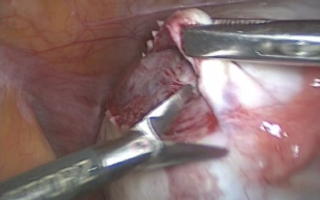
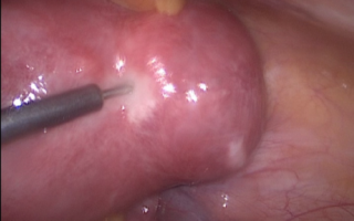
Locate fibroid and inject inj. Vasopressin in particular dilution with saline . This injection helps in reducing blood flow to fibroid ( by constricting the vessels supplying fibroid ). This is needed as fibroid is very vascular organ .

Step 2

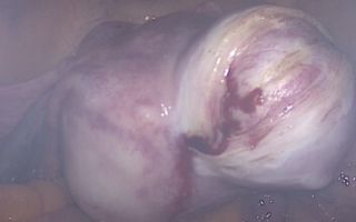
Taking incision on fibroid depending on type of fibroid.

Step 3

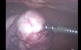
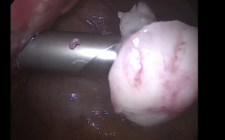
Inserting a screw like device ( myoma screw ) in fibroid to take out the fibroid from its capsule inside the uterus

Step 4

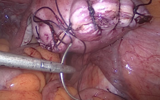
Suturing of uterus at the site from where myoma is removed ( myoma bed ) with absorbable suture material in 2 -3 layers

Step 5

Removing the myoma outside the abdomen by use of special instrument called as morcellator. It uses mechanical energy to make small pieces of fibroids in the form of strips an then taken out of the abdomen through same incision.

What do you mean by diagnostic laparoscopy and operative laparoscopy for infertility?

Diagnostic laparoscopy for infertility is performed as a part of investigation for infertility in cases where no abnormality is detected on non invasive tests like USG (ultrasound) or HSG (hysterosalpingography). It is usually performed in women with unexplained infertility, following failure of repeated IUIs or along with operative hysteroscopy. The pelvic anatomy is visualized with a telescope like instrument. The uterus, tubes, ovaries and there surrounding structures are systematically checked. A blue coloured dye is injected from the cervix (mouth of the uterus) by an assistant and the dye is seen coming out through the fallopian tubes via the telescope, thus confirming that the tubes are open. This is known as chromopertubation.